The Slippers Were Faster Than I Expected
The ammonia test strips have been reading 2.0 ppm for three days. The bacterial bloom hasn’t cleared. Yesterday morning, squinting at the test kit’s little colour chart, I noticed something moving in the sample water I’d just pipetted out of the tank.
Not debris. Not bubbles. Something with intent.
I dug out the stereoscope from my electronics bench — the one I use for inspecting solder joints on QFP packages — and put together a crude wet mount. Drop of tank water on a glass slide, coverslip lowered at an angle to push the air bubbles out. Under 20× magnification: paramecia. Dozens of them, spiralling through the murk like tiny slippers propelled by invisible oars.
The cloudy aquarium water I’d been cursing as a failure indicator wasn’t just chemistry. It was an ecosystem I couldn’t see.
A stereoscope is the wrong tool for this. The magnification tops out around 40×, and what I was looking at deserved more. By midnight I’d ordered a compound microscope — an AmScope B120C, nothing fancy, but it has a mechanical stage and Abbe condenser, which apparently matter. It arrived this afternoon.
Setting up a compound microscope properly involves something called Köhler illumination, a technique from 1893 that I’d never heard of despite decades of using optical instruments. The principle is counterintuitive: you adjust two diaphragms — the field diaphragm near the lamp and the condenser diaphragm below the stage — to deliberately defocus the light source. The goal is to make the lamp filament invisible, producing even illumination across the specimen. Without it, you get hot spots and shadows that masquerade as specimen features.
It took forty minutes to get this right. The manual’s instructions assumed I knew what a “field diaphragm” was and where to find it. I didn’t. Trial and error, adjusting rings until the image quality suddenly improved.
Second wet mount of the day, this time at 100×. The paramecia weren’t just visible — they were fast. At this magnification, their movement across the field of view is startling, like watching time-lapse traffic from a helicopter. They travel at roughly a millimetre per second in real life, which doesn’t sound impressive until you see it through a lens that makes them fill half your visual field.
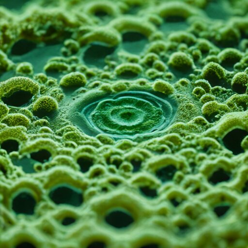
Louis Joblot, a French microscopist, first described these in 1718 and called them “chaussons” — slippers. The name persisted for two centuries. Looking at them now, the resemblance is obvious: a rounded toe, a slight heel, the whole shape elongated like something you’d shuffle around the house in. They rotate as they move, corkscrewing through the water, and the cilia covering their surface are just barely visible as a shimmering haze.
I stayed at 100× for most of the afternoon. Higher magnification exists — this scope goes to 1000× with the oil immersion objective — but increasing power shrinks your field of view and demands thinner specimens. At 400×, I could see individual bacteria in the water, tiny rods darting between the larger organisms, presumably the heterotrophic bloom that’s been making the tank look like milk. The bacterial cloud I wanted gone is actually the foundation of everything else I was watching. Food chain, visible in a droplet.
The wet mount dried out after twenty minutes. This is apparently standard — you’re watching on borrowed time. I made three more before dinner, each from a different part of the tank. The substrate water near the aquasoil had the most activity. The surface film had diatoms, golden-brown rectangles that don’t move but cluster together in chains. The filter outflow had almost nothing, which makes sense — that water has been through mechanical filtration.
One observation I can’t explain: some paramecia were noticeably larger than others, maybe 1.5× the body length, and they moved more slowly. Different species? Different life stage? The identification guides I’ve found online require staining to distinguish species, and staining kills the organism. I’d rather keep watching than categorize.
There’s a version of this hobby that involves prepared slides — permanent mounts of professionally sectioned specimens, stained and preserved, catalogued by tissue type. Educational, precise, archival. The local scientific supply company sells sets of fifty for reasonable prices. I’ll probably order some eventually, for the days when I want to understand histology rather than just observe movement.
But tonight, the tank water. The Hemianthus is still melting — maybe half the original planting has yellowed and collapsed — but under the microscope, the system that’s killing those plants is also sustaining something. Rotifers, I think, though I’m not certain. Transparent organisms with wheel-like structures at one end, anchored to debris, spinning. They showed up in the second wet mount and were gone by the third.
Antonie van Leeuwenhoek, the cloth merchant who accidentally invented microbiology in the 1670s, called these things “animalcules.” He wrote chaotic letters to the Royal Society describing what he saw through hand-ground lenses no better than the cheap stereoscope I started with this morning. He was elected Fellow of the Royal Society despite having no scientific training, purely because he’d looked more carefully than anyone else at water that everyone assumed was empty.
The tank is still cloudy. The ammonia is still elevated. But the numbers on the test strips are less interesting to me now than what’s swimming between them.